Pathophysiological role of free reactive iron species in hypoxic tissues during the development of organ failure
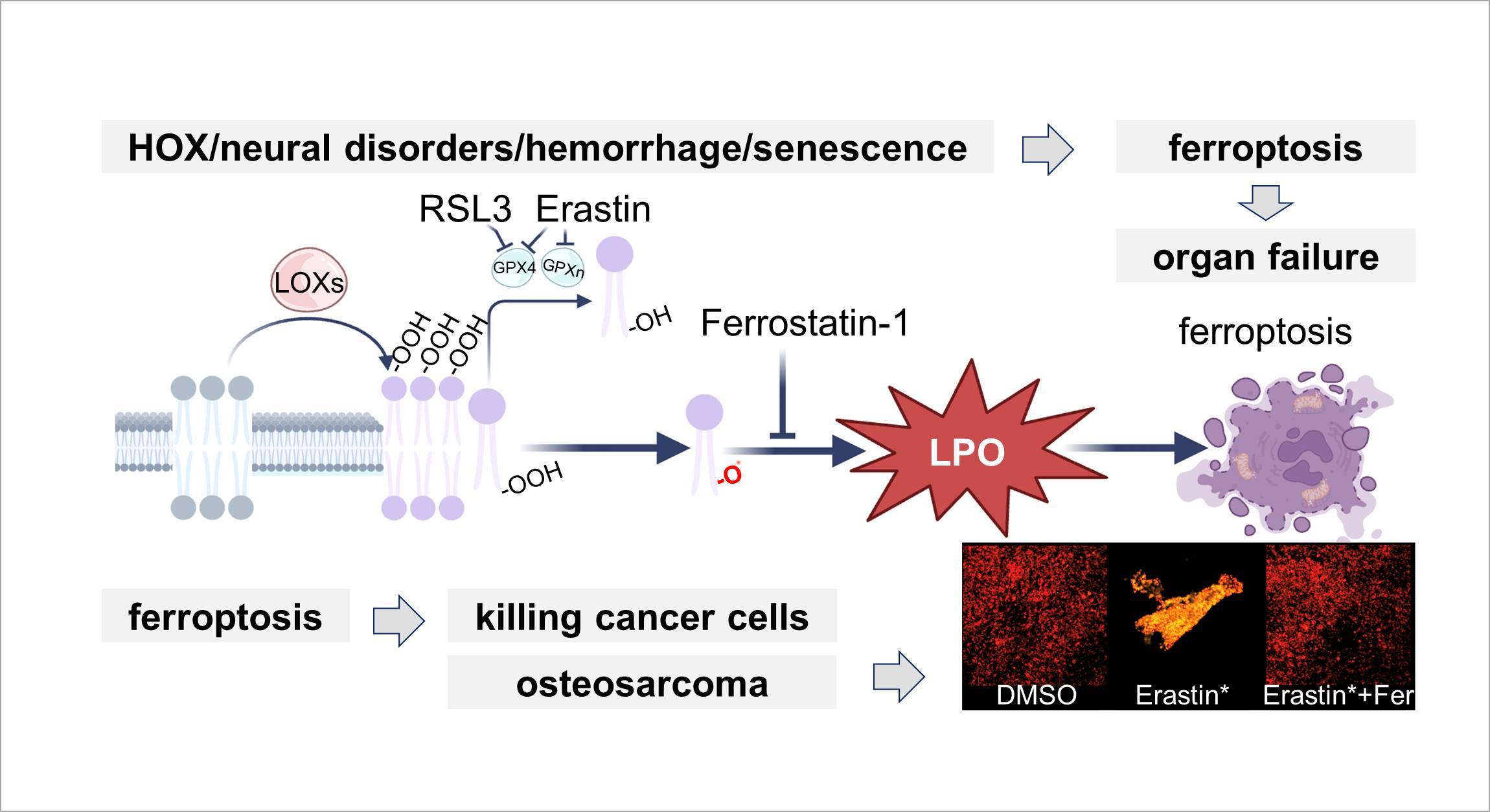
A number of studies in recent years provided evidences that hypoxia, neural disorders, hemorrhage and senescence induce ferroptosis, a recently discovered programmed cell death. Ferroptosis is an iron-dependent, non-apoptotic cell death, which can lead to organ failure. The formation of cellular lipid hydroperoxides (LOOH) is catalyzed by iron-dependent enzyme, lipoxygenases (LOX). Under normal conditions, the LOOH are removed from the cellular metabolism by specific enzymes, glutathione peroxidases (GPXs). Ferroptosis is characterized by a reduction in intracellular glutathione and decreased activity of GPXs, which prevents the detoxification of LOOH. Ferroptosis can be induced by pharmacological inhibition of GPXs, for example with RSL3 or Erastin. Ferrostatin-1, a synthetic antioxidant, acts via a reductive mechanism to prevent damage to membrane lipids and thereby specifically inhibits ferroptosis. Fluorescent images demonstrate a series of reduced and oxidized BODIPY-C11 probe signals with a drastic increase in lipid peroxidation (LPO) in ferroptosis. It is shown that osteosarcoma cells are very sensitive to ferroptosis, suggesting that the induction of ferroptosis is a potential tool to combat osteosarcoma.