Cartilage Regeneration

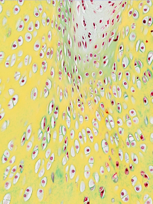
Articular cartilage defects are caused by multiple reasons such as damage, abuse or genetic predisposition but have in common that they do not heal. They cause pain and require clinical intervention at a certain stage. Especially sports injuries are the reason for movement problems of young patients which particularly require regeneration rather than a replacement of damaged tissue by artificial materials such as metal, ceramics or plastic. Osteoarthritis is prevalent in elderly people and requires a systematic approach including treatment of the surrounding tissues and inflammation.

The group around Sylvia Nürnberger develops systems to grow cartilage in order to replace degraded or damaged cartilage and non-functional repair tissue. Currently, the most promising approach is the use of biomaterials operating as scaffold materials for cells. The scaffolds are either seeded with the cells in vitro before implantation or are implanted simultaneously with the cells during surgery. Research focuses on:

  • Application of decellularized tissue as scaffolds for allogenic application
  • Development of methods to repopulate the decellularized cartilage tissue with cells
  • Engineering of scaffold materials with controlled parameters (design, mechanical properties and composition)
  • Studying substances with potential positive effect on cell fitness and chondrogenesis

The overall aim is to improve and accelerate the regeneration process of the current cell-based cartilage treatment strategies and extend the limits of defect size towards large surface treatment. Furthermore, the functionality of scaffolds with adipose derived stromal cells (ASCs) as an alternative cell source to autologous chondrocytes is investigated. The application of the amniotic membrane as a gliding tissue is part of the research done in the area of tendon regeneration.

a. Cartilage cells in their lacunae within dense extracellular matrix.

Selected Publications

Schneider C, Nürnberger S (2023). Decellularization of Articular Cartilage: A Hydrochloric Acid-Based Strategy. Methods Mol Biol. 2023;2598:301-311. 

Baranovskii D, Demner J, Nürnberger S, Lyundup A, Redl H, Hilpert M, Pigeot S, Krasheninnikov M, Krasilnikova O, Klabukov I, Parshin V, Martin I, Lardinois D, Barbero A (2022). Engineering of Tracheal Grafts Based on Recellularization of Laser-Engraved Human Airway Cartilage Substrates. Cartilage. 2022 Jan-Mar;13(1):19476035221075951. 
(free PDF)

Hölzl K, Fürsatz M, Göcerler H, Schädl B, Žigon-Branc S, Markovic M, Gahleitner C, Van Hoorick J, Van Vlierberghe S, Baudis S, Pauschitz A, Redl H, Ovsianikov A, Nürnberger S (2022). Gelatin methacryloyl as environment for chondrocytes and cell delivery to superficial cartilage defects. J Tissue Eng Regen Med. 2022 Feb;16(2):207-222.

Feichtinger X, Heimel P, Tangl S, Keibl C, Nürnberger S, Schanda JE, Hercher D, Kocijan R, Redl H, Grillari J, Fialka C, Mittermayr R (2022). Improved biomechanics in experimental chronic rotator cuff repair after shockwaves is not reflected by bone microarchitecture. PLoS One. 2022 Jan 5;17(1):e0262294.
(free PDF)

Fürsatz M, Gerges P, Wolbank S, Nürnberger S (2021). Autonomous spheroid formation by culture plate compartmentation. Biofabrication. 2021 Jan 29.

Nürnberger S, Schneider C, Keibl C, Schädl B, Heimel P, Monforte X, Teuschl AH, Nalbach M, Thurner PJ, Grillari J, Redl H, Wolbank S (2021). Repopulation of decellularised articular cartilage by laser-based matrix engraving. EBioMedicine. 2021 Jan 16;103196.

Bachmann B, Spitz S, Schädl B, Teuschl AH, Redl H, Nürnberger S, Ertl P (2020). Stiffness Matters: Fine-Tuned Hydrogel Elasticity Alters Chondrogenic Redifferentiation. Front Bioeng Biotechnol. 2020 Apr 30;8:373.
(free PDF)

Nürnberger S, Schneider C, van Osch GVM, Keibl C, Rieder B, Monforte X, Teuschl AH, Mühleder S, Holnthoner W, Schädl B, Gahleitner C, Redl H, Wolbank S (2019). Repopulation of an auricular cartilage scaffold, AuriScaff, perforated with an enzyme combination. Acta Biomater. 2019 Mar 1;86:207-222.

Rieder B, Weihs AM, Weidinger A, Szwarc D, Nürnberger S, Redl H, Rünzler D, Huber-Gries C, Teuschl AH (2018). Hydrostatic pressure-generated reactive oxygen species induce osteoarthritic conditions in cartilage pellet cultures. Sci Rep. Nov 19;8(1):17010.
(free PDF)

Nürnberger S, Rentenberger C, Thiel K, Schädl B, Grunwald I, Ponomarev I, Marlovits St, Mejer Ch & Barnewitz D (2017). Giant crystals inside mitochondria of equine chondrocytes. Histochem Cell Biol. 147(5):635-649
(free PDF)

Schneider C, Lehmann J, van Osch GJVM, Hildner F, Teuschl A, Monforte X, Miosga D, Heimel P, Priglinger E, Redl H, Wolbank S & Nürnberger S (2016). Systematic comparison of protocols for the preparation of human articular cartilage for use as scaffold. Tissue Engineering Part C, 22(12):1095-1107.
(author’s manuscript)

Utomo L, Mieke M, Pleumeekers, Nimeskern L, Nürnberger S, Stok K, Hildner F & van Osch G (2015). Preparation and characterization of a decellularized cartilage scaffold for ear cartilage reconstruction. Biomedical Materials, 10(1):015010.

Lindenmair A, Nürnberger S, Stadler G, Meinl A, Hackl C, Eibl J, Gabriel C, Hennerbichler S, Redl H & Wolbank S (2014). Intact human amniotic membrane differentiated towards the chondrogenic lineage. Cell Tissue Bank, 15(2):213-225.

Nürnberger S, Barnewitz D, Tichy B, Ponomarev I, Meyer C, Klepal W, Albrecht C & Marlovits S (2013). (Ultra-)Structural details of cells and matrix during cartilage regeneration and differentiation in MACT-treated defects in the horse. Journal of Tissue Science and Engineering, S2:1-10.
(free PDF)

Teuschl AH, Nürnberger S, Redl H & Nau T (2013). Articular cartilage tissue regeneration: current research strategies and outlook for the future. European Surgery, 45(3):142-53.

Albrecht Ch, Tichy B, Hosiner St, Nürnberger S, Zak L, Aldrian S & Marlovits S (2013). Influence of cryopreservation, cultivation time and patient’s age on the gene expression in cartilage transplants. International Orthopaedics, 37(11):2297-2303.
(free PDF)

Nürnberger S, Meyer C, Ponomarev I, Barnewitz D, Resinger C, Klepal W, Albrecht Ch & Marlovits S (2013). Equine Articular Chondrocytes on MACT Scaffolds for Cartilage Defect Treatment. Anatomia, Histologia, Embryologia: Journal of Veterinary Medicine Series C, 42(5):332-343.

Albrecht Ch, Tichy B, Nürnberger S, Hosiner S, Zak L, Aldrian S & Marlovits S (2011). Gene expression and cell differentiation in matrix-associated chondrocytes transplantation grafts: a comparative study. Osteoarthritis and Cartilage, 19 (10):1219-1227.
(free PDF)

Nuernberger S, Cyran N, Albrecht Ch, Redl H, Vécsei V & Marlovits S (2011). The influence of scaffold architecture on chondrocyte distribution and behavior in matrix associated chondrocyte transplantation grafts. Biomaterials, 32(4):1032-1040.

Hildner F, Peterbauer A, Wolbank S, Nürnberger S, Marlovits S, Redl H, van Griensven M & Gabriel C (2010). FGF-2 abolishes the chondrogenic effect of combined BMP-6 and TGF-b in human adipose derived stem cells. J Biomed Mater Res A, 94(3):978-987.

Nürnberger S, Tichy B, Jabersberger T, Vécsei V & Marlovits S (2006). Histological Aspects of Articular Cartilage. Osteosynthesis and Trauma Care, 14(3):158-167.

Nürnberger S, Klepal W, Vécsei V & Marlovits S (2006). Ultrastructural Insights into the World of Cartilage – Electron Microscopy of articular cartilage. Osteosynthesis and Trauma Care, 14(3):168-180.