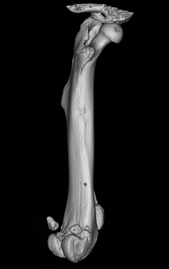
Broken bones, ruptured ligaments or injuries of inner organs but also damages on a cellular level are often a consequence of trauma. To follow healing processes and visualize the basic mechanisms of regeneration, various imaging techniques are used. This encompasses different methods that create imaging material on various levels, from organs to cellular structures.
The group from Paul Slezak established a core facility for pre-clinical imaging techniques that includes for example optical methods, X-ray and CT-imaging as well as magnetic resonance and experimental imaging. Hereby, various aspects of the research can be captured visually and long-term profiles of regeneration can be created. The following methods are used:
- In vivo fluorescence
- Bioluminescence
- µ- and nano-CT
- Laser-Doppler imaging
- Optoacustic imaging
These imaging techniques can for example trace the formation and regeneration of tissues such as cartilage and bones. Mechanisms of vascularization can be made visible by imaging and the degradation of biomaterials or therapeutic materials can be tracked. Furthermore, the distribution of active substances in the organism can be made visible in order to assess for example the efficiency of the methods of administration.