Opening up new perspectives
“Please do not lean” is written on a small paper card folded on a buzzing grey box. Wrinkles in the paper tell us that many a person has nevertheless leaned on the box. The box is inconspicuous, and yet the micro-CT is one of the most important and expensive instruments at the Ludwig Boltzmann Institute for Traumatology (“LBI Trauma”). The research institute in cooperation with AUVA has one of the best devices in the country in use.
Micro-CT or micro-computed tomography makes it possible to view the body in completely new dimensions. Similar to CT machines in clinical practice, where an X-ray machine rotates around the body part, micro-CT takes X-ray images from all angles. These are then put together in the computer to form a 3D image. What makes the micro-CT device so special is its high resolution: the resolution goes down to the nanometre range, i.e. less than a thousandth of a millimetre. A big advantage is that the sample is not destroyed by the measurement. It does not have to be cut into slices, as is the case with histology. With a literal X-ray view, the device looks inside bones and teeth. To see softer structures such as vessels or nerves well, it needs a little more assistance.
Spinal cord research
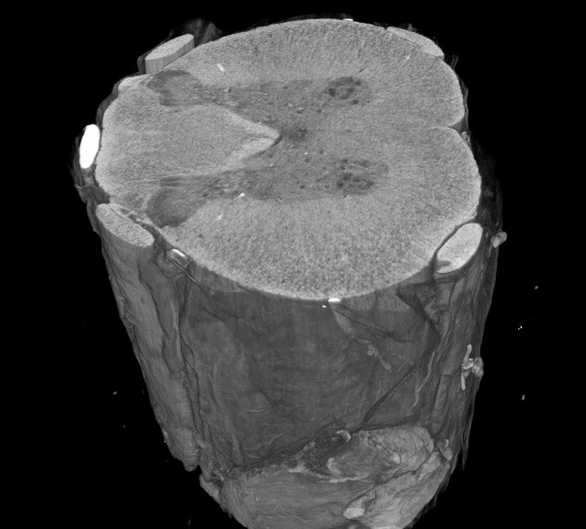
The “blind spot” of soft tissue can be eliminated in CT by using contrast media. Modern CT techniques provide unprecedented insights, for example into nerve tissue. At LBI Trauma, an old contrast agent method, Lugol’s staining, named after its discoverer, was recently rediscovered and optimised for combination with micro-CT measurement. The technique is now in use in spinal cord research. Scientists can now see down to the smallest nerve fibres and cells. This is invaluable in the search for treatment options for spinal cord contusions.
Measuring “bucky balls”
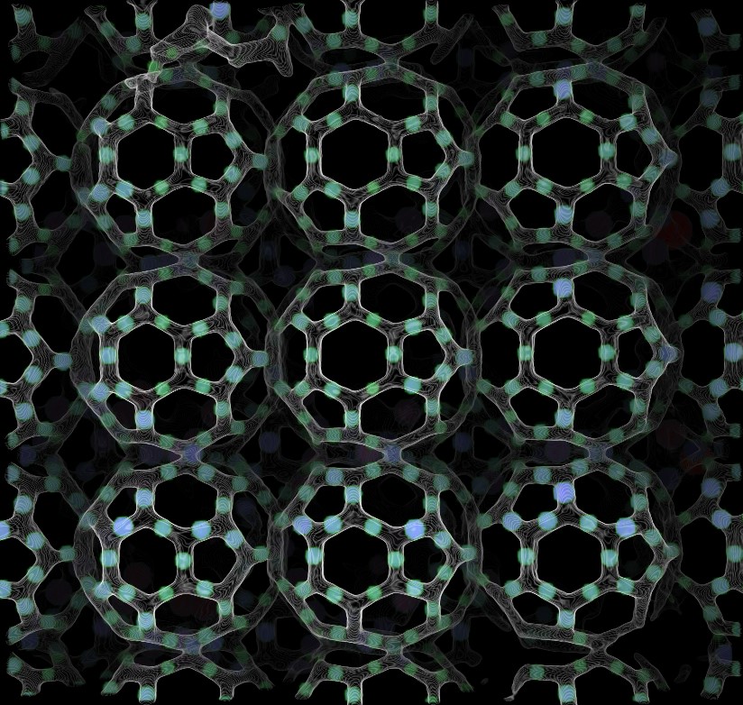
In the laboratory of Prof. Dr. Aleksandr Ovsianikov at the Vienna University of Technology, the so-called “bucky balls” were developed. The microscopically small scaffolds, reminiscent of footballs, come from the 2-photon 3D printer and have a diameter of only 300 micrometres. Three pieces lined up fit into a millimetre. The micro-CT impressively illustrates the fine, regular framework structures and that the “bucky balls” can be joined together to form large structures. The small balls can adapt to different defect shapes as an implant and allow cells to grow in to form new body tissue.
Scanning bones
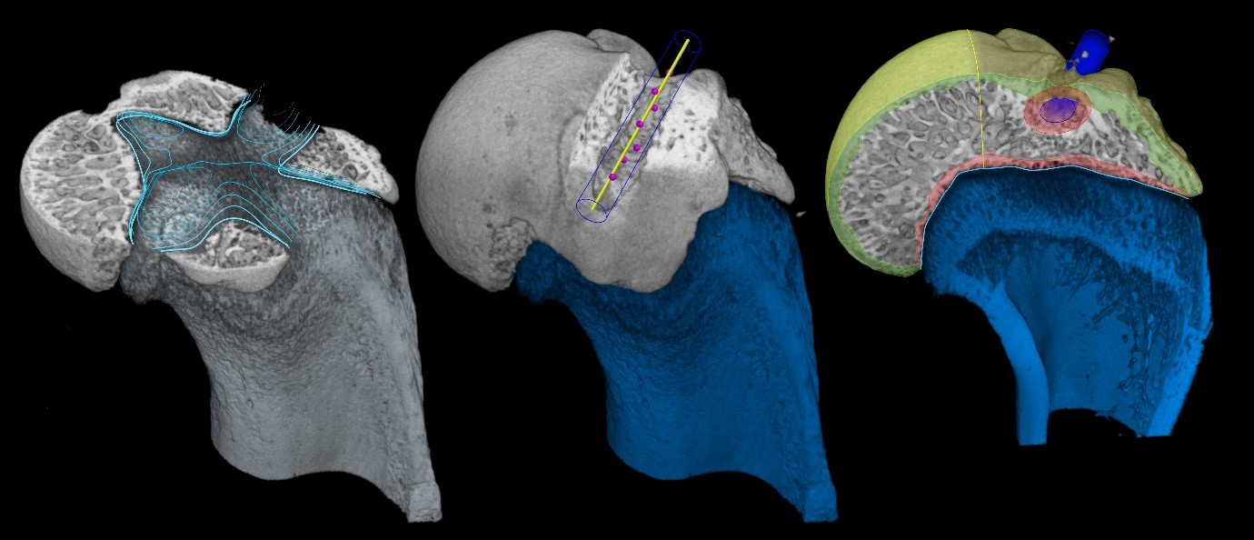
Measuring bones is a home game for micro-CT. The bioinformatician at LBI Trauma, Patrick Heimel, MSc, has been involved in over 50 studies on the topic of bones alone. Particularly prominent is the study by Dr. Jakob Schanda, in which the team was able to prove that the administration of an osteoporosis drug after a complex tendon rupture in the shoulder increases the bone density in the shoulder head. Typical bioinformatics methods were used: the CT scans were converted into formats suitable for measurement and the different bone regions – the thick outer shell and the inner, spongy framework – were marked in the software. A wide variety of parameters can then be calculated in the computer. Of particular interest in the study were the structure and density of the inner scaffolding of the shoulder head, as its condition often deteriorates after a shoulder tendon injury. This is where the drug investigated in the study showed an effect. The article published on this subject has won several awards.
Use in archaeology
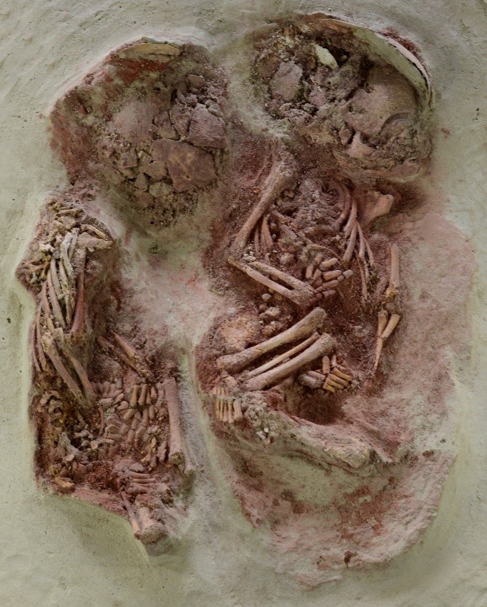
Hard tissue is not only highly interesting in medicine, it also attracts a lot of attention in archaeology – after all, bones and teeth are usually the only things left over from ancient bodies. Patrick Heimel has also been allowed to participate in studies in cooperation with the Natural History Museum, under the direction of Maria Teschler-Nicola, Director of the Anthropological Department. Together with the anthropologist Stefan Tangl and the imaging specialist Toni Dobsak from the University Dental Clinic in Vienna, he dealt with the question of the exact age at death of the “Wachtberg twins”, two infants found in an excavation site in Lower Austria, who died about 31,000 years ago. Using microCT, they succeeded in making the so-called neonatal line visible in the tooth enamel. In the micro-CT, this stands out as a dark line in the enamel and separates the prenatally from the postnatally formed enamel. This made it clear that one twin outlived the other by about six weeks. The twins are thus simultaneously the youngest and oldest “patients” ever examined in the micro-CT of the LBI Trauma.